
ZEISS Apotome Plus
Precision Redefined in Fluorescence Microscopy- 00 năm
- 00 tháng
- 00 ngày
- 00 giờ
- 00 phút
- 00 giây
 imaged with Plan Apochromat 63x/1.4.")
 imaged with Plan Apochromat 63x/1.4.")
Reliable Optical Sections
Under Varying Experimental ConditionsApotome 3 significantly increases the axial resolution compared to conventional fluorescence widefield microscopy: You obtain optical sections that allow 3D rendering, even from thick specimens. Three grids of different geometries give you the best resolution for each objective. You can focus on your experiment as the ideal illumination structure is automatically selected, always resulting in high-contrast optical sections.
Caption: Cos7 cells (nuclei stained with Hoechst, tubulin with Alexa 488 and Phalloidin with Alexa 568) imaged with Plan Apochromat 63x/1.4.
")
")
")
")
Peer-reviewed Algorithms
Linear Approaches for True Optical SectionsPurely software-based methods require either prior knowledge of the sample (AI based methods) or rely on complex algorithms that have not been peer-reviewed. Users must trust that these black-box solutions do not falsify information when “enhancing” the image. ZEISS Apotome 3 uses the information from the structured illumination combined with documented algorithms to create a crisp optical section you can trust.
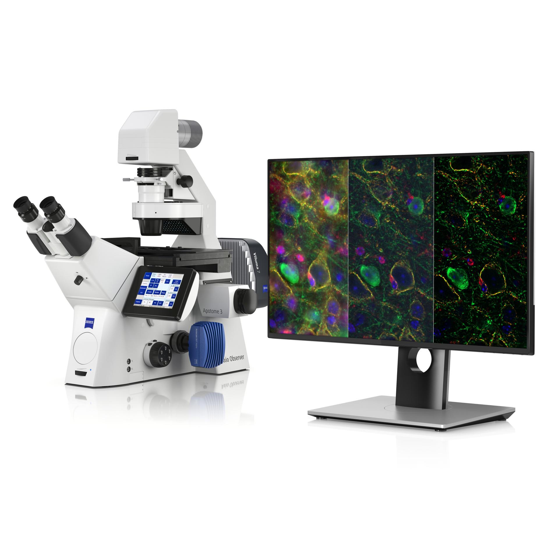
Caption: Cortical neurons (left: Widefield; right: Apotome 3). Courtesy of L. Behrendt, Leibniz-Institute on Aging – Fritz-Lipmann-Institut e.V. (FLI), Germany.


Sagittal Section of Adult Mouse Brain
Confocal-like Image Quality
180 nm Resolution with Apotome PlusResolve details that were not visible before with your widefield microscope: With Apotome Plus, you can obtain structural information at lateral resolution down to 180 nm. The combination of structured illumination with state-of-the-art image processing substantially improves the signal-to-noise ratio and resolution in x, y, and z.
Caption: 35 μm sagittal section of adult mouse brain, imaged with ZEISS Axio Observer and ZEISS Apotome, processed with Apotome Plus. Sample courtesy of University of California, Davis / NIH NeuroMab Facility.
Agenda
-
Unlocking Precision in Fluorescence Microscopy with ZEISS Apotome Plus
09:00 – 09:30
Registration and Welcome Coffee
Arrive, network, and collect materials.
09:30 – 09:45
Welcome Address
Introduction to ZOYC and the importance of advancing microscopy techniques.09:45 – 10:30
Keynote: Structured Illumination in Fluorescence Microscopy
Understand the science behind structured illumination and how it achieves optical sectioning.10:30 – 11:15
Session: ZEISS Apotome Plus – The System and Its Applications
Learn how Apotome Plus works, its capabilities, and its relevance in biological research.11:15 – 11:30
Break
Refreshments and informal networking.11:30 – 12:15
Case Studies: From Cells to Complex Samples
Explore imaging examples, including live-cell imaging, thick tissue sections, and 3D renderings.12:15 – 13:00
Live Demo: Imaging with ZEISS Apotome Plus
See a real-time demonstration of optical sectioning and high-resolution imaging.13:00 – 14:00
Lunch Break
Enjoy lunch and connect with peers and ZEISS experts.14:00 – 14:45
Interactive Workshop: Optimising Sample Preparation and Imaging
Learn best practices for sample preparation and hands-on tips for optimal imaging.14:45 – 15:30
Future Perspectives in Widefield Microscopy
Discuss emerging trends and how ZEISS supports evolving research needs.15:30 – 16:00
Q&A and Closing Remarks
Ask questions and recap key takeaways.16:00 – 17:00
Networking and Meet the Experts
Engage directly with ZEISS specialists and explore additional resources. -
Hands-on demonstration - Test your own specimen
Register for an individual hands-on demonstration and test your own specimen using the Lattice SIM 3 or Celldiscoverer 7. Participation in the sample preparation presentation is strongly recommended for a successful demonstration.
Select your demonstration in the form
Gentle super-resolution live cell imaging with ZEISS Lattice SIM 3
Adaptable automation for advanced workflows with ZEISS Celldiscoverer 7


